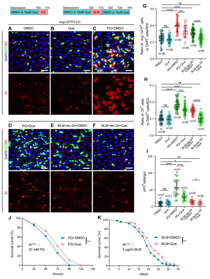
Figure 2.
Que prevents stimulus-induced hyperproliferation of ISCs and improves stress tolerance in Drosophila. (A–F) Representative immunofluorescence images of Drosophila posterior midguts stained with DAPI, GFP, and Dl for DMSO (A), Que (B), PQ + DMSO (C), PQ + Que (D), BLM-rec-2d + DMSO (E), and BLM-rec-2d + Que (F) groups. The 0-day-old flies were fed with 10 µM Que for 10 days, then treated with 10 mM PQ for 1 day, followed by dissection. The 0-day-old flies were fed with 10 µM Que for 10 days, then treated with 25 µg/mL BLM for 1 day, and then resumed feeding with 10 µm Que for 1/2/3 days, followed by dissection. The top panels represent the merged images, whereas the bottom panels represent Dl. Scale bars represent 25 µm. (G,H) The ratio of esg-GFP+ cells (G) and Dl+ cells (H) to DAPI+ cells per ROI in the posterior midguts of flies in experiments (A–F). n: number of ROI counted. (I) The number of pH3+ cells in the whole gut of flies in experiments (A–F). n: number of gut counted. (J,K) Survival percentage of female W1118 flies with DMSO (blue curve) or Que (pink curve) supplementation under 20 mM PQ (J) or 5 µg/mL BLM (K) treatments. Each group had 100 flies and three independent experiments were conducted. Error bars represent SDs. Log-rank test was used for lifespan analysis. Student’s t-tests, * p < 0.05, ** p < 0.01, *** p < 0.001, **** p < 0.0001, and non-significance (ns) represents p > 0.05.

