Figure 4.
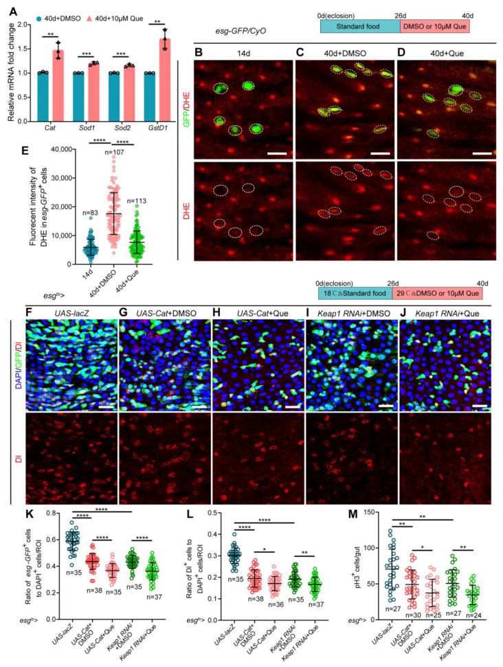
Que prevents age-related hyperproliferation of ISC partially through its antioxidative ability. (A) RT-qPCR analysis of the antioxidant-related genes (Cat, Sod1, Sod2, and GstD1) in the midguts of 40-day-old flies fed with or without Que. Three independent experiments were conducted. (B–D) Representative immunofluorescence images of the posterior midguts of 14- (B) and 40-day-old (C) flies without Que supplementation and 40-day-old flies with 10 µM Que supplementation (D) stained with DHE (red; ROS maker). Esg-GFP+ cells and DHE staining are circled by the white dashed line. The top panels represent the merged images, whereas the bottom panels represent DHE staining. Scale bars represent 10 µm. (E) Quantitation of DHE fluorescence intensity in esg-GFP+ cells from experiments (B–D). Each dot indicates one esg-GFP+ cell. (F–J) Representative immunofluorescence images of posterior midguts of flies carrying esgts-Gal4-driven UAS-lacZ (F), UAS-Cat + DMSO (G), UAS-Cat + Que (H), Keap1 RNAi + DMSO (I), and Keap1 RNAi + Que (J) stained with DAPI, GFP, and Dl. The top panels represent the merged images, whereas the bottom panels represent Dl. Scale bars represent 25 µm. (K,L) The ratio of esg-GFP+ cells (K) and Dl+ cells (L) to DAPI+ cells per ROI in the posterior midguts of flies in experiments (F–J). n: number of ROI counted. (M) The number of pH3+ cells in the whole gut of flies in experiments (F–J). n: number of gut counted. Error bars represent SDs. Student’s t-tests, * p < 0.05, ** p < 0.01, *** p < 0.001, and **** p < 0.0001.

