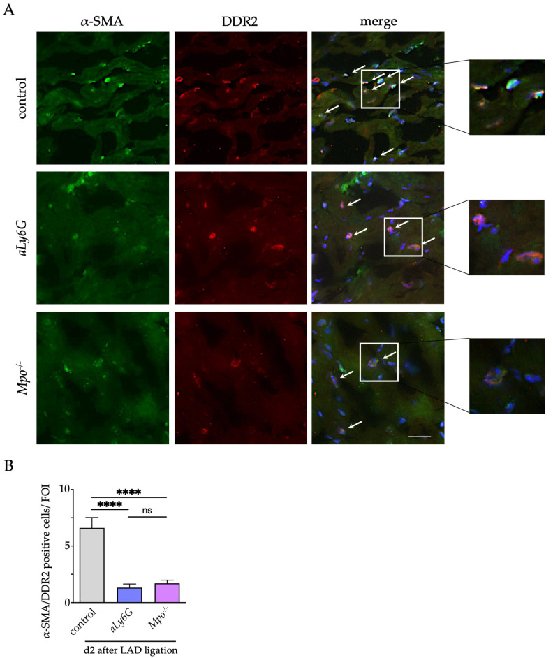
Figure 4.
Myofibroblast transdifferentiation after MI. (A) Representative immunofluorescence stainings of murine cardiac sections 2 days after PI for the fibroblast marker discoidin domain-containing receptor 2 (DDR-2; green) and the myofibroblast marker α–smooth muscle actin (α-SMA; red) (blue = DAPI; scale bar = 100 μm). Arrows indicate double positive cells. (B) Quantification of myofibroblasts within the infarct and peri-infarct region. n = 5/5/5; **** p < 0.0001, ns = not significant; one way ANOVA test followed by appropriate post hoc test, mean ± SEM is shown.

