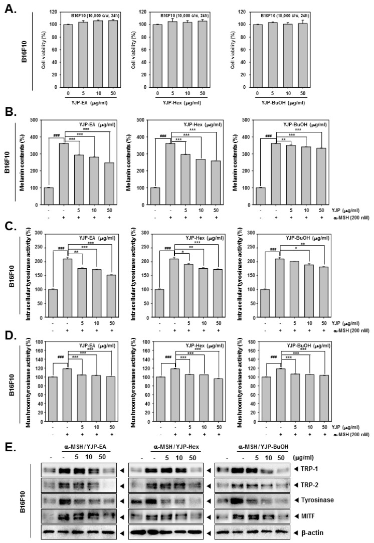
Figure 4.
Melanin inhibition effects of YJP-EA, Hex, and BuOH on B16F10 cells. (A) B16F10 cells were treated with YJP-EA, Hex, and BuOH for 24 h. Next, the cell viability was measured using the MTT assay. (B) Melanin contents from α-MSH-stimulated B16F10 cell. The cells were treated with α-MSH (200 nM) and YJP-EA, Hex, and BuOH for 48 h. The cell lysates were measured at 490 nm. (C) To measure intracellular tyrosinase activity, the B16F10 cells were treated with -MSH (200 nM) and YJP-EA, Hex, and BuOH for 72 h. L-DOPA was added into the lysates and measured at 490 nm. (D) The mushroom tyrosinase activity was evaluated at -MSH (200 nM) and in YJP-EA-, Hex-, and BuOH-treated B16F10 cells. The supernatants with L-DOPA and tyrosinase were measured at 475 nm. (E) The whole cell lysates were analyzed by Western blot analysis. All experiments were performed individually in triplicate. *** p < 0.001 vs. α-MSH-treated cells, ** p < 0.01 vs. α-MSH-treated cells, and * p < 0.05 vs. α-MSH-treated cells. ### p < 0.001 vs. non-treated (NT) cells.

