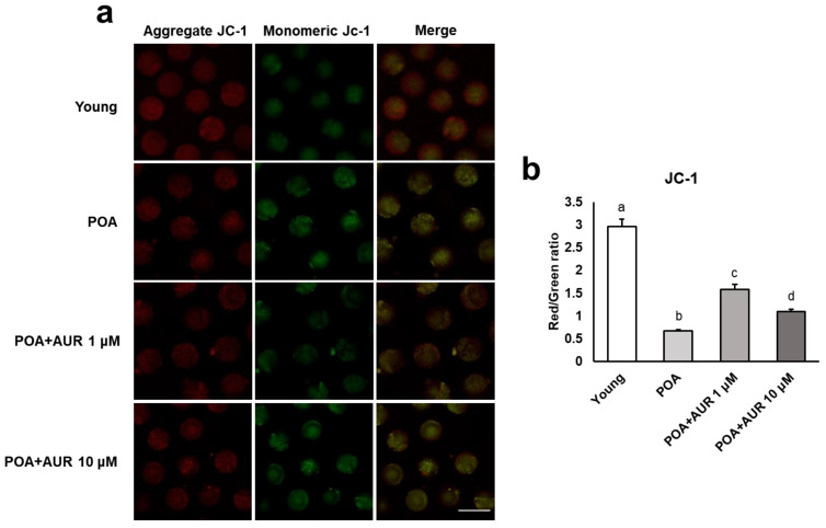
Figure 3.
AUR enhanced mitochondrial function by raising ΔΨm during postovulatory aging. (a) Representative images of mitochondrial ΔΨm in young and aged oocytes with or without AUR administration. Oocytes were stained with the cationic dye JC-1, which can selectively enter mitochondria and reversibly turn the emitted green fluorescence into red depending upon the mitochondrial membrane potential (aggregated JC-1: high potential marker, red; monomeric JC-1: low potential marker, green); scale bar = 20 μm. (b) The ratio of red to green fluorescence intensity in each group of oocytes. Data are presented as the means ± SEMs of at least three independent experiments. Significant difference indicated by lowercase letters of the alphabet, p < 0.001.

