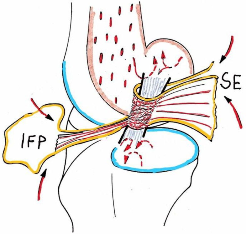
Figure 1.
Schematic drawing of the blood supply to the canine cranial cruciate ligament; IFP, infrapatellar fat pad, SE, synovial envelope (epiligament): arrows indicate afferent supply, broken arrows show endosteal vessels, only marginally (from proximal) or not entering ligament matrix (from Niebauer GW, Pathomechanisms in canine cruciate ligament rupture <in German>. PhD Thesis 1982, Vet. Med Univ. Vienna, Austria).

