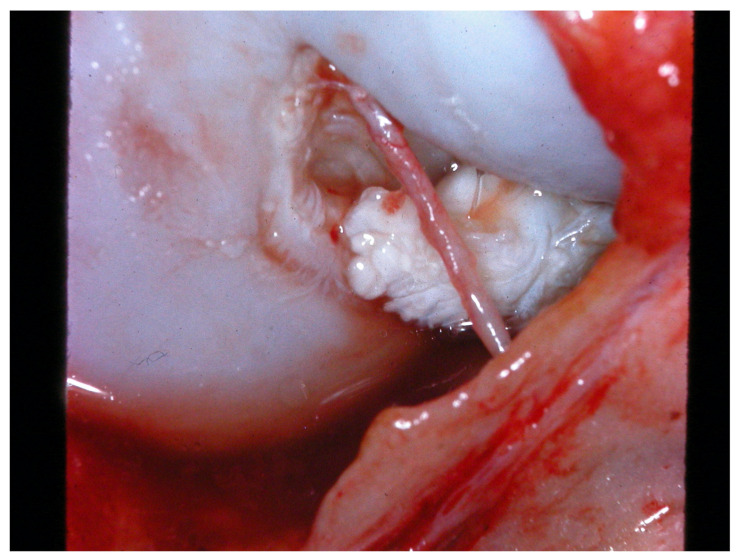
Figure 2.
Intra-operative image of a cranial cruciate ligament ruptured 4 weeks previously; note the distal ligament stump with rounded fibrillar edges due to ongoing collagenolysis; surface is partly covered by an inflammatory tissue membrane (reddish patches). The structure visible in front of the ligament is the remnant of the epiligamentous synovial shield which covered the intact cruciate ligament (image by the author).

