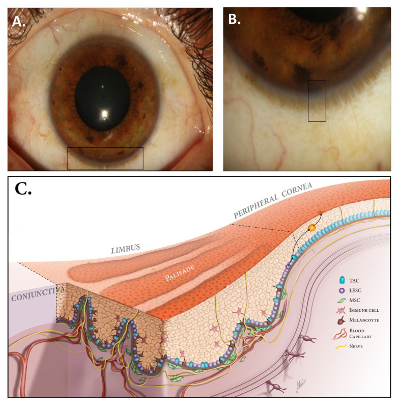
Figure 1.
Normal ocular surface and limbus. (A) The corneoscleral limbus contains the palisades of Vogt (PVs), which have a length of 0.31 mm and a width of 0.04 mm and are typically more detectable on the superior and inferior sections of cornea. (B) Corneoscleral junction with magnification showing PVs. (C) The PVs contain different cells, such as melanocytes, mesenchymal stem cells, and immune cells. These cells, along with neurovasculature, provide growth factors, nutrients, and structural support to promote proper LESC proliferation and differentiation (LESC: limbal epithelial stem cell, TAC: transient amplifying cell, MSC: mesenchymal stem cell). Modified with permission from [14].

