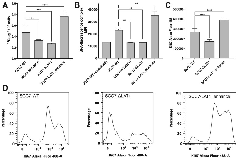
Fig. 2.
p-Boronophenylalanine (BPA) uptake capacity based on the expression of LAT1 in cancer cells. (A) Boron concentration in tumor cells with genetically modified tumor cells assessed via inductively coupled plasma atomic emission spectrometry (n = 6). Values were normalized as 10B μg per 107 cells. (B) MFI of the complex of BPA and BPA-probe in tumor cells evaluated via flow cytometer (n = 4). (C) Mean Alexa Fluor 488 fluorescent intensity of Ki67 evaluated via flow cytometer (n = 5). (D) Representative histogram of the Ki67 fluorescent intensity of SCC7-WT, SCC7-ΔLAT1 and SCC7-LAT1_enhance. P-values of < 0.05 were considered to be statistically significant (**P < 0.01, ***P < 0.001, ****P < 0.0001).

