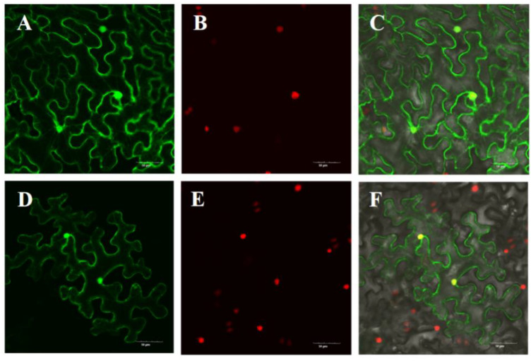
Figure 7.
Subcellular localization of LsOVATE1 in N. benthamiana epidermal cells. Empty GFP vectors were used as controls, and showed green fluorescence. The tobacco nuclei showed red fluorescence. C and F are digitally merged images of both bright-field and fluorescent signals. (A–C) show the localization of empty GFP control vector; (D–F) show the localization of LsOVATE1. Scale bar = 50 µM.

