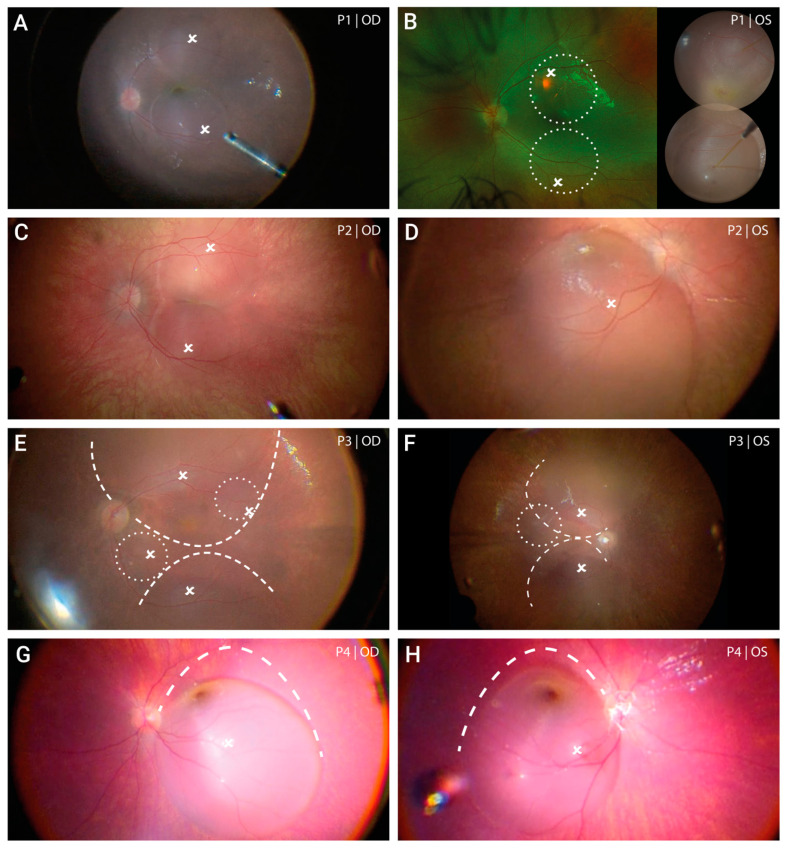
Figure 1.
Extent of subretinal gene therapy with voretigene neparvovec. In both the right (A) and the left eye (B) of patient 1 two subretinal blebs were formed of which the first bleb placed at the superior vascular arch led to foveal detachment. Due to limited intraoperative records, the final extent of the subretinal bleb formation of the left eye is schematically shown by white circles placed on a pre-operative fundus image, and is supplemented by an image of the two blebs during injection. Treatment of the right eye in patient 2 (C) consisted of two subretinal blebs that merged at the level of the fovea (kissing blebs). In the left eye (D) one large bleb was formed with marked spread to the superior periphery. Subretinal injection in patient 3 was challenging due to a remarkable shift of the vector solution towards the retinal periphery. After initial injection, two smaller blebs (white circles) were added in the right (E) and one bleb in the left eye (F). In both eyes, treatment of the posterior pole could be achieved by enlarging the bleb through gentle suction at the margins of the bleb using a backflush flute needle. This procedure was performed following fluid-air-exchange. The extent of enlargement is illustrated by the dashed lines. Both the right eye (G) and left eye (H) of patient 4 was treated with one large bleb. Following injection, the bleb was enlarged to maximize the treated area using the same technique as described for patient 3. Please note: all intraoperative images (except for P1 OS) are inverted and laterally reversed (surgeon’s view). White crosses indicate the subretinal injection sites. Dashed lines indicate the final edges of the bleb after enlargement.

