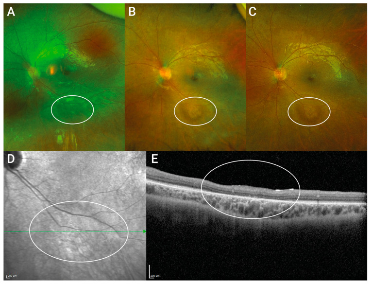
Figure 2.
Photoreceptor layer loss at the injection site. Upper Panel shows postoperative wide-angle fundus images of P1 taken 4 weeks (A), 23 months (B) and 32 months (C) after subretinal gene therapy. The roundish irregular lesion at the injection site at the inferior vascular arcade was recognized first 4 weeks after therapy and did not extend over time. (D) near-infrared scanning laser ophthalmoscope fundus image 32 months after therapy shows only mild changes. (E) OCT through the lesion taken 32 months after gene therapy revealed circumscribed photoreceptor layer loss at the injection site but no atrophy of the choroid nor the RPE was observed.

