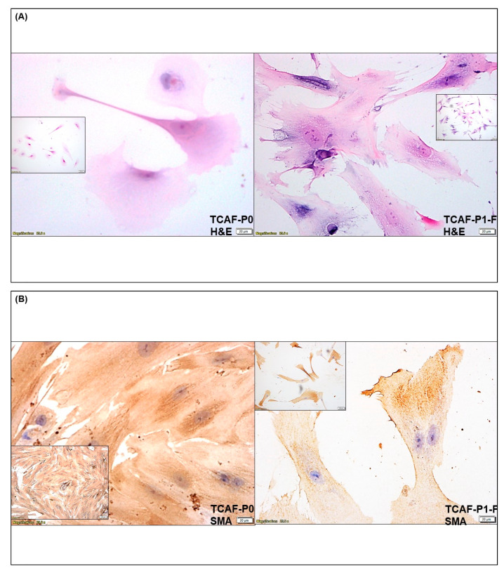
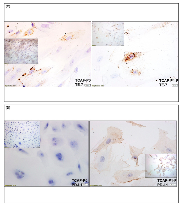
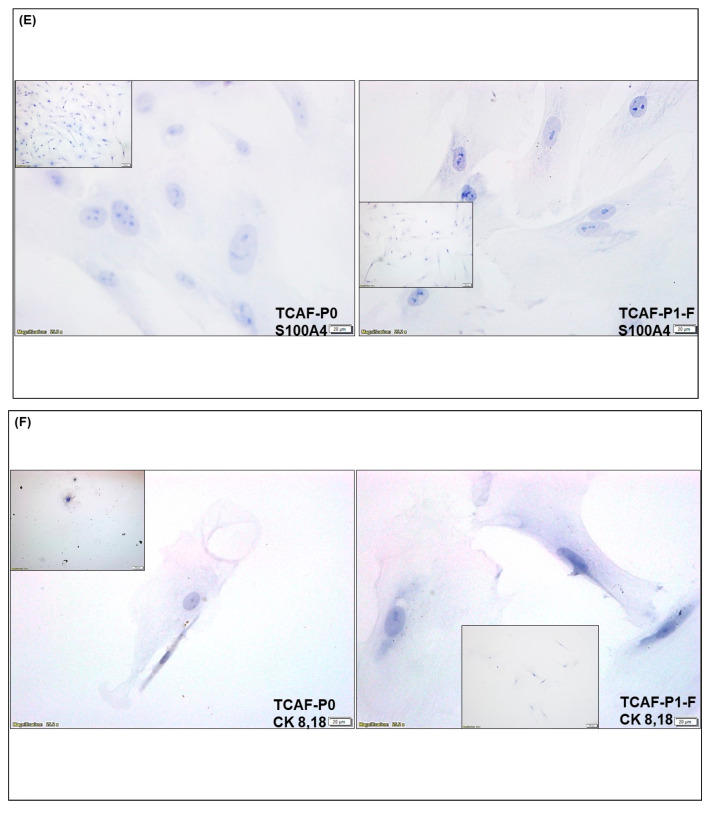
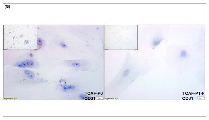
Figure 5.
Cellular localization of proteins for CAF markers in the Patient-Derived primary culture of ovarian CAF from pre- and post-frozen passages established from the representative tumor sample, HU-A148-Ov2 of ovarian cancers: We tested the cellular localization of the expression of protein by ICC for SMA (B), TE-7 (C), PD-L1 (D), S100A4 (E), CK 8, 18 (F), and CD31 (G) in Patient-Derived CAFs (before and after thawing). The morphological features are presented by H&E stain (A). Insets represent photomicrographs in lower magnification.


