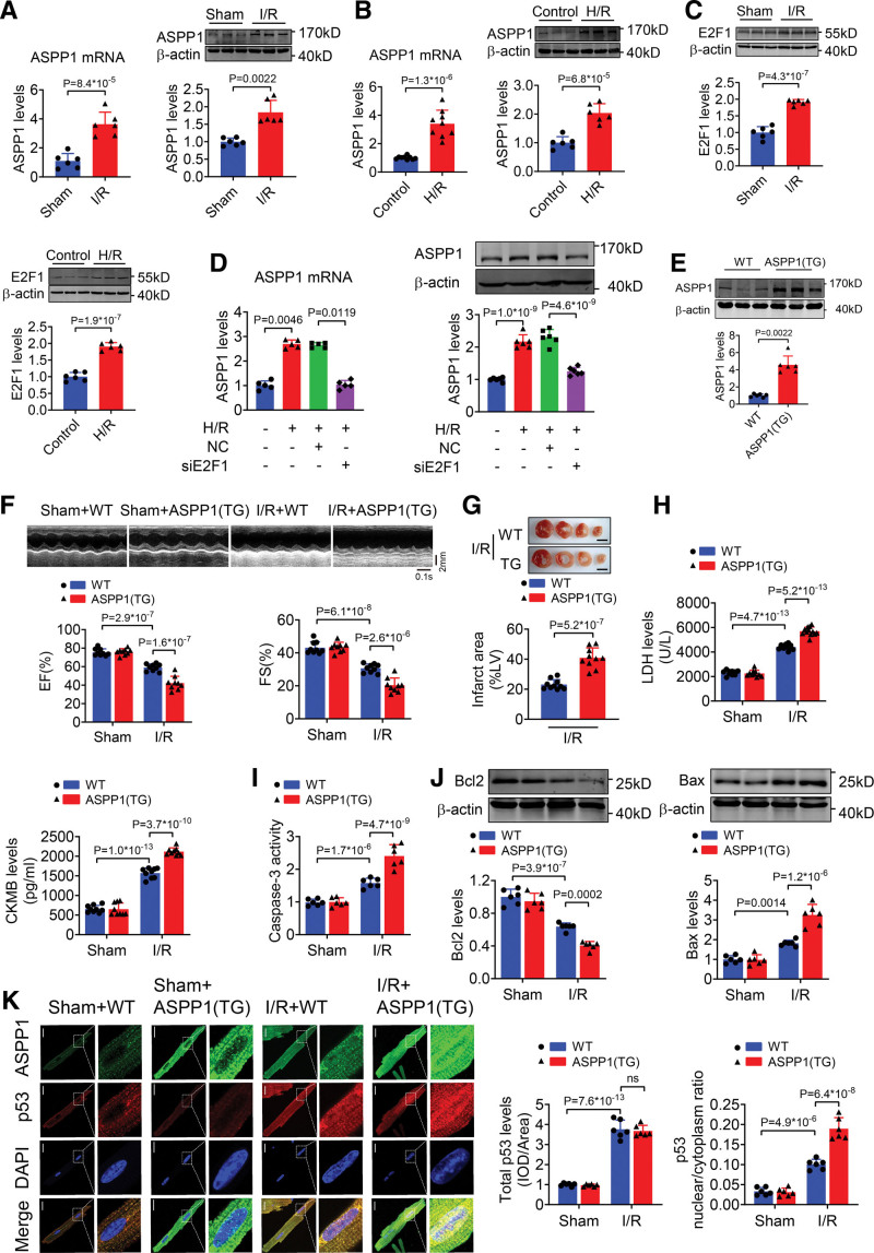
Figure 3.
Cardiomyocyte-specific overexpression of ASPP1 aggravates cardiac I/R injury. A, The mRNA (Student t test) and protein (Mann-Whitney U test) levels of ASPP1 in cardiac tissues of mice subjected to 45 minutes ischemia followed by 24 hours reperfusion (I/R); n=6. B, The mRNA and protein levels of ASPP1 in neonatal mouse ventricular cardiomyocytes (NMVCs) subjected to H/R (Student t test); n=9 for mRNA level; n=6 for protein level. C, The protein levels of E2F1 in I/R hearts and NMVCs treated with H/R stimulation by Western blot (Student t test); n=6. D, Transfection of E2F1 siRNA inhibited the mRNA (Kruskal-Wallis, followed by false discovery rate [FDR] method of Benjamini and Hochberg test) and protein (1-way ANOVA, followed by Tukey post hoc multicomparisons test) expression of ASPP1 in NMCMs exposed to H/R by quantitative real-time PCR (qRT-PCR) and Western blot assay; n=5 for mRNA level; n=6 for protein level. E, The protein levels of ASPP1 in wild type (WT) and ASPP1 transgenic mice by Western blot (Mann-Whitney U test); n=6. F, Cardiac function of wild type (WT) and ASPP1 transgene mice after I/R injury by echocardiography (2-way ANOVA, followed by Tukey post hoc multicomparisons test); n=9. G, Infarct area of I/R mice by 2,3,5-triphenyltetrazolium chloride (TTC) staining (Student t test); n=10. Scale bar=2 mm. H, Serum LDH and creatine kinase isoenzyme MB (CKMB) levels (2-way ANOVA, followed by Tukey post hoc multicomparisons test); n=10 (WT+Sham and ASPP1 transgene+Sham); n=12 (WT+IR and ASPP1 transgene+I/R) for LDH assay; n=9 for CKMB assay. I, Caspase-3 activity detected by ELISA assay (2-way ANOVA, followed by Tukey post hoc multicomparisons test); n=9. J, The protein levels of Bcl2 and Bax detected by Western blot (2-way ANOVA, followed by Tukey post hoc multicomparisons test); n=6. K, Immunofluorescent staining was performed to analyze the effect of ASPP1 transgene on p53 nuclear translocation in isolated adult cardiomyocytes after cardiac I/R injury (2-way ANOVA, followed by Tukey post hoc multicomparisons test); n=6. ns, not significant. Scale bar=20 μm. ASPP1 indicates apoptosis stimulating of p53 protein 1; DAPI, 4′,6-diamidino-2-phenylindole; EF, ejection fraction; FS, fractional shortening; H/R, hypoxia/reoxygenation; and I/R, ischemia/reperfusion.

