Figure 4.
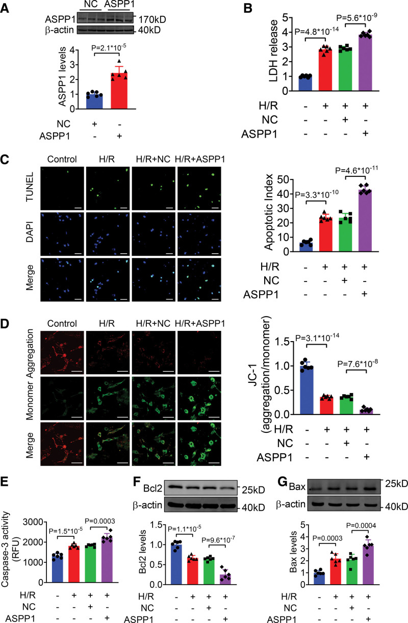
Overexpression of ASPP1 aggravates H/R-induced damage in neonatal mouse ventricular cardiomyocytes (NMVCs). A, The efficiency of ASPP1 overexpression plasmid in NMVCs by Western blot (Student t test); n=6. B, LDH release (1-way ANOVA, followed by Tukey post hoc multicomparisons test); n=6. C, Apoptosis of NMVCs detected by TUNEL (TdT-mediated dUTP nick end labeling) assay (1-way ANOVA, followed by Tukey post hoc multicomparisons test); n=6. Scale bar=50 μm. Green, TUNEL -positive cells; blue, DAPI. D, Mitochondrial membrane potential of NMVCs by JC-1 assay (1-way ANOVA, followed by Tukey post hoc multicomparisons test); n=6. Scale bar=50 μm. E, Caspase-3 activity in NMVCs by ELISA assay (1-way ANOVA, followed by Tukey post hoc multicomparisons test); n=6. F and G, The protein levels of Bcl2 (F) and Bax (G) detected by Western blot (1-way ANOVA, followed by Tukey post hoc multicomparisons test); n=6. ASPP1 indicates apoptosis stimulating of p53 protein 1; Bax, Bcl2-associated X; DAPI, 4′,6-diamidino-2-phenylindole; H/R, hypoxia/reoxygenation; and LDH, lactate dehydrogenase.

