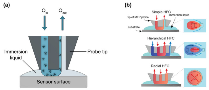
Figure 21.
(a) Illustration of simple microfluidic printing (µFP) probe used to pattern a sensor surface with a bioreceptor solution (antibody solution illustrated here as an example). (b) Comparison of µFP using simple hydrodynamic flow confinement (HFC), hierarchical HFC, which permits recirculation of the patterning solution in the µFP head, and radial HFC, which produces circular, rather than teardrop-shaped spots. The processing (bioreceptor) solution is shown in red, the immersion liquid is shown in light blue, and the shaping liquid used for HFC is shown in dark blue. Insets on the right show the printing footprints for each µFP type. Part (b) is adapted with permission from Ref. [32]. Copyright 2021 American Chemical Society.

