Figure 1.
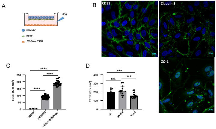
The in vitro blood–brain barrier (BBB) model. (A) Scheme of porcine brain microvascular endothelial cells (PBMVEC), human brain microvascular pericytes (HBVP) and SV40 large T-antigene immortalized astrocytic cells (SV-GA) or T98G glioblastoma cell line localization in the BBB co-culture model co-culture. (B) CD31, claudin 5 and ZO-1 staining of isolated PBMVEC (bars = 10 µm). (C) The cells were cultivated as described in the material and methods part either as monocultures (HBVP on the bottom insert membrane; PBMVEC on the top insert membrane) or as co-cultures. After 5 days, the membrane integrity was determined by TEER measurement (n = 3, each up to 10 replicates, SEM, t-test, **** p < 0.0001). (D) Co-culture of barrier-tight BBB-layers (containing PBMVEC and HBVP) with SV-GA or T98G cells. TEER was performed 24 h after co-culture (Co: no additional cells were seeded; n > 7, SEM, t-test, *** p < 0.005). Co-culture with astrocytic cells strengthens, with glioma cells weakens barrier integrity. (C,D) Circles, squares and triangles at the appropriate bars indicate data points of single experiments.

