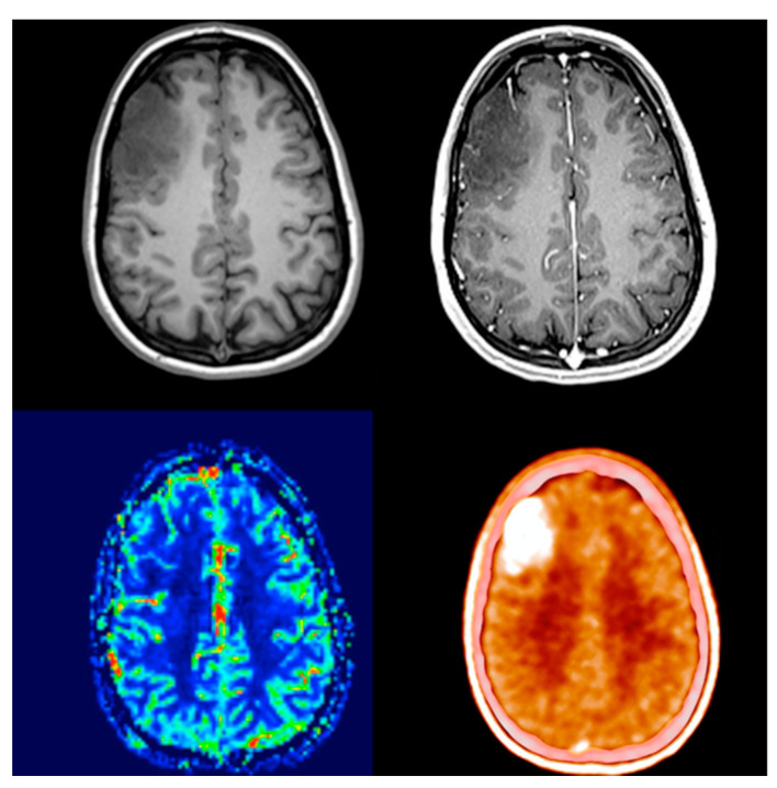
Figure 1.
Thirty-two-year-old female with a right frontal non-enhancing mass known since the age of 14. Presenting for follow-up examination after 18 years of absence. The size of the lesion showed significant progression. Either contrast enhancement or elevated blood flow was present. PET/CT scan showed a smaller region and intense, well-circumscribed, nearly homogenous amino acid uptake (SUVmax: 4.95 vs. 1.57; TBRmax: 3.17). After surgical removal, histology revealed anaplastic oligodendroglioma with IDH mutation and 1p/19q co-deletion.

