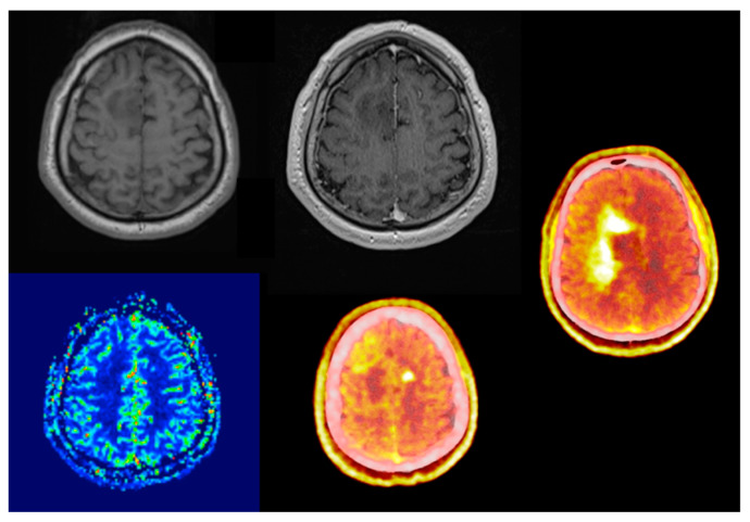
Figure 2.
Sixty-three-year-old male, right frontal non-enhancing lesion known for over a year. During follow-up MRI scans no radiological progression was detectable. There were also no signs of elevated regional blood flow perfusion. No neurological deficit upon first and following presentations. PET/CT scan presented a butterfly-like very intense heterogeneous, lesion, significantly widened compared to the MRI lesion. Bilateral amino acid uptake (SUVmax: 55.81 vs. 1.58; TBRmax: 3.68) with a photopenic (SUV 1.4) area in the T1 hypointense area in the lateral part of the MRI lesion. The original T2 hyperintense lesion was photopenic (SUV: 1.4). Stereotactic biopsy revealed IDH-positive astrocytoma (grade 4), Ki-67 proliferation rate was 40%.

