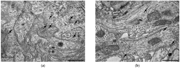
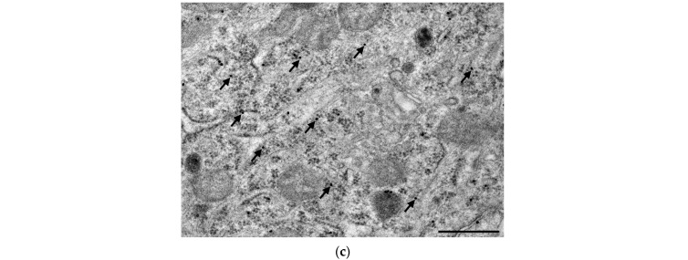
Figure 6.
Representative electron micrographs of myenteric neuronal perikaryon and ganglionic neuropil from the ileum of a control rat after TLR4 post-embedding immunohistochemistry. The number of TLR4 labelling gold particles was higher in neuronal perikaryon (a,c) than in myenteric neuropil (a,b). Arrows—18 nm gold particles labelling TLR4, scale bars: 500 nm.


