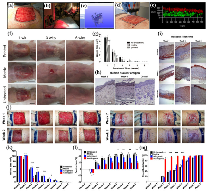
Figure 6.
In situ bioprinting of autologous skin cells for healing of full-thickness wounds. (a) The full-thickness wound on the porcine skin. (b) Geometric information was obtained by 3D scanner. (c) Scanned data with its coordinate system. (d) In situ printing process. (e) The layering of FBs (green) and KCs (red) in the skin constructs. (f) Observation of wound healing on the mouse model using printed, dressing matrix, and untreated control for 6 weeks. (g) Size of the wound for 6 weeks. (h) Anti-human nucleus shows that human cells were maintained for 6 weeks. (i) Masson’s trichome staining showed fast re-epithelialization in the printed group compared to the dressing matrix and untreated control. (j) Observation of wound healing process in the porcine model. (k) Wound size, (l) contraction, and (m) re-epithelialization, during 8 weeks, show that the printed skins containing autologous cells induced excellent wound healing. The asterisks (*~***) refer to significant differences (* = p < 0.05; ** = p ≤ 0.01, and *** = p ≤ 0.001). The data were reproduced from Ref [105] Copyrights 2018 Nature Publishing Group.

