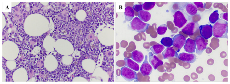
Figure 2.
Bone marrow morphology in a representative case (case #8). (A) Bone marrow core biopsy shows a hypercellular marrow with increased immature cells as well as many dysplastic megakaryocytes with small hypolobated or separated nuclear lobes (100×); (B) Bone marrow smear shows increased blasts that are large with dispersed chromatin and distinct nucleoli (500×).

