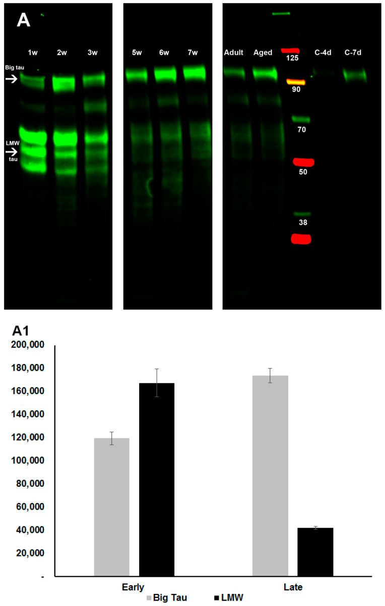
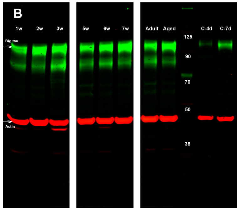
Figure 3.
Developmental regulation of Big tau expression in SCG. Western blot analysis of SCG tissue prepared from different ages including 1–3, and 5–7 postnatal weeks, adult at 4 months, and aged animals at 12 months, as well as dissociated SCG cultures from week-5 postnatally cultured for 4 and 7 days. Samples were stained with the 3′tau antibodies, which recognize all tau isoforms as described in Methods. The blot (A) shows that the LMW (45–60 kDa) tau isoforms are present at postnatal weeks 1–3 of SCG together with Big tau at 110 kDa. The levels of the LMW tau isoforms gradually decreased until Big tau became the dominant isoform at 5–7 postnatal weeks as well as in adult and aged animals. (A1) shows the quantitative analysis of band densities, depicting the relative levels of Big tau and LMW tau at early postnatal stages (average of weeks 1–3) and late postnatal stages (average of weeks 5–7) using raw data corrected relative to actin shown in (B) (e.g., the 2 w lane was overloaded and the C-4d underloaded). A parallel set of Western blots with identical samples were stained with Big tau antibodies (B) confirming that the expression of Big tau in SCG increased during early postnatal stages and remained the dominant isoform in SCG in adult and aged animals, as well as cultured dissociated SCG neurons. C-4d, C-7d: dissociated SCG neurons from week-5 that were cultured for 4 and 7 days, respectively. The blots included a pre-stained protein ladder and were probed for expression of actin (42 kDa). The levels of actin were used to normalize loading variations in assessment of changes in the level of expression of the different tau isoforms (shown in (B) as red bands and used for both (A,B)).


