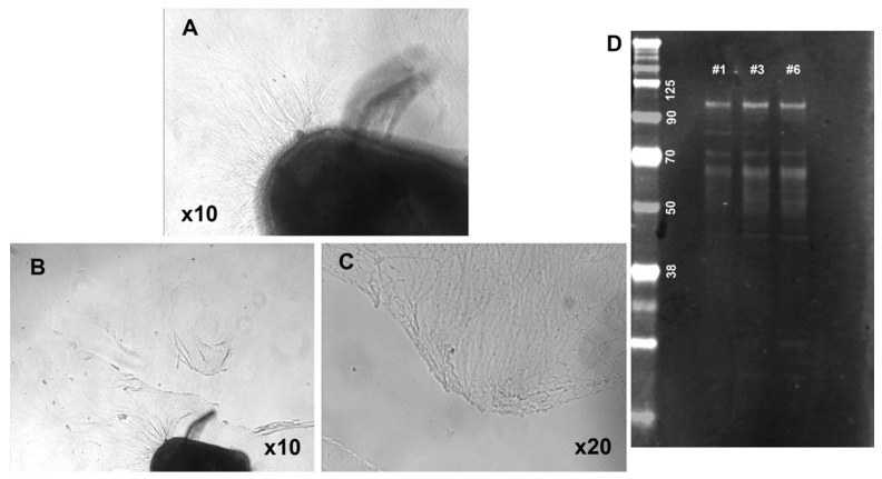
Figure 8.
Characterization of SCG explants (P3) cultured for 8 days and lesioned. Phase contrast images show the network of neurites growing around the explant (A). They were lesioned at about half of their length (B,C) and shown at low (B) and high (C) magnification immediately after the lesion, then allowed to regrow (Figure 9 and Figure 10). Western blot stained with the 3′ tau antibodies show Big tau expression at 110 kDa (D) in the P3 explants grown for 8 days, which were lesioned and cultured overnight (lanes #1 and #3) with lane #6 showing the explant without the lesion.

