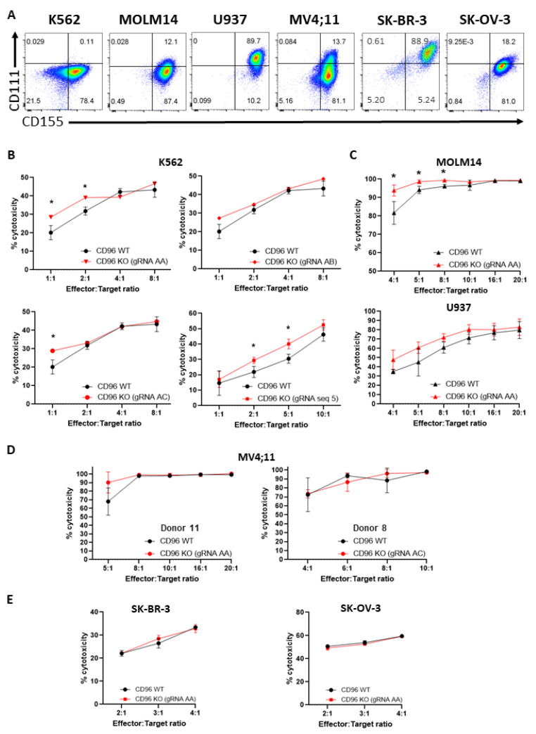
Figure 2.
CD96 deficiency in T cells enhances T cell cytotoxicity against K562 and MOLM14 cells. (A) Expression of CD96 ligands, CD155 and CD111, on K562, MOLM14, U937, MV4;11, SK-BR-3, and SK-OV-3 cells as assessed by flow cytometry. Quadrant gates were applied based on no staining for CD155 and CD111. (B) Percentage (%) cytotoxicity of T cells (calculated as described in Materials and Methods) against luciferase-expressing K562 cells 20 h following co-incubation of T and K562 cells at indicated E:T ratios. Each graph depicts T cells that were electroporated with different gRNAs. (C) % cytotoxicity of T cells against luciferase-expressing MOLM14 (top) and U937 (bottom) co-cultured with T cells as in (B). (D) % cytotoxicity of T cells, derived from 2 different human donors, against luciferase-expressing MV4;11 cells co-cultured with T cells as in (B). (E) % cytotoxicity of T cells against luciferase-expressing SK-BR-3 (left) and SK-OV-3 (right) co-cultured with T cells as in (B). Data in (B–E) are the mean ± SD of 3 technical replicates; multiple unpaired Student’s t-tests, *, p < 0.05. All data are representative of 2 independent experiments.

