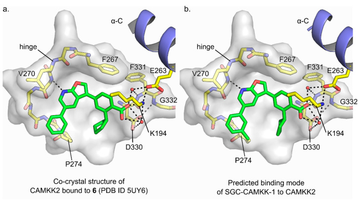
Figure 4.
Predicted binding mode of probe SGC-CAMKK1 to CAMKK2 kinase domain. (a) Detailed view of the binding interactions between compound 6, a close furopyridine analog of probe SGC-CAMKK-1, and CAMKK2 ATP-binding pocket as seen in the complex co-crystal structure (PDB ID 5UY6). (b) Predicted binding mode of probe SGC-CAMKK1 to CAMKK2 ATP-binding pocket obtained by in silico docking (see Methods for details). In (a,b), dashed black lines indicate potential hydrogen bonds, water molecules are depicted as red spheres, carbon atoms are shown in green (ligand) or yellow (protein), the kinase domain α-helix C is shown in blue, and protein surface (for the bottom of the ATP-binding site) is shown in white.

