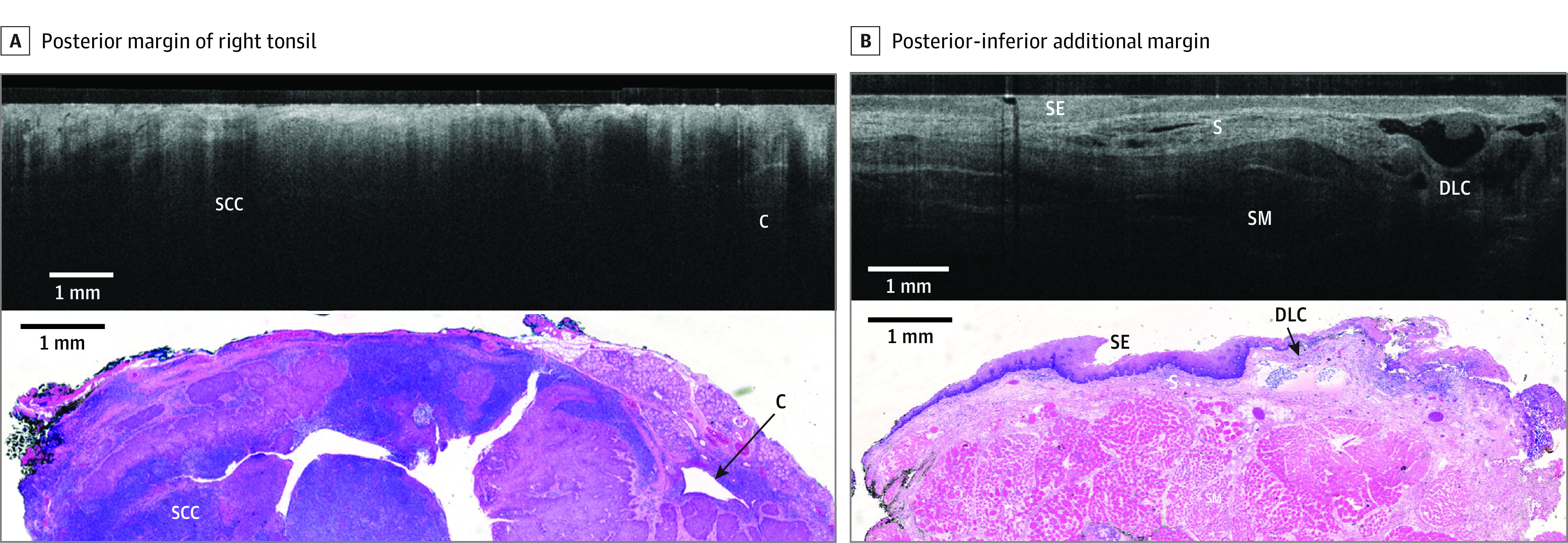
Figure 2. Representative Comparison of Images for Patient A.

Comparison is shown between wide-field optical coherence tomography and permanent histology of invasive, moderately differentiated squamous cell carcinoma (SCC, p16 negative) with lymphatic invasion. A, The posterior margin of the right tonsil is shown, with wide-field optical coherence tomography in the top panel showing SCC as an area with decreased light penetrance depth compared with the adjacent normal tonsillar lymphatic tissue. In the bottom panel, a hematoxylin and eosin slide from the corresponding region is shown. A crypt (C) is also seen on the right side of the image. B, The posterior-inferior additional margin is shown, with wide-field optical coherence tomography in the top panel showing squamous epithelium (SE), submucosal layer (S), and skeletal muscle (SM). In the bottom panel, a hematoxylin and eosin slide from corresponding region is shown. A dilated lymphatic channel (DLC) is also seen on the right side of the image.
