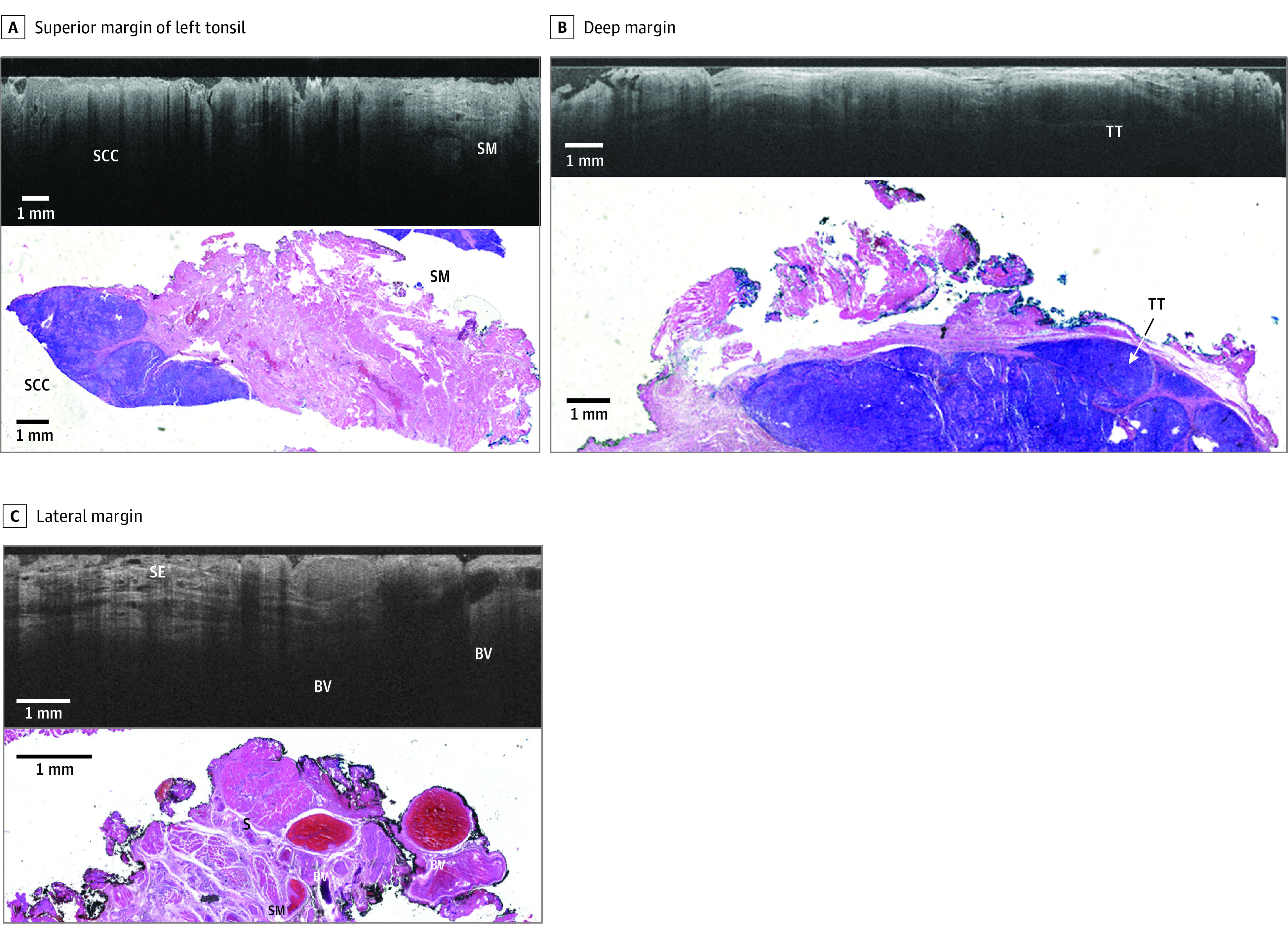
Figure 3. Representative Comparison of Images for Patient B.

Comparison is shown between wide-field optical coherence tomography and permanent histology of human papillomavirus–related squamous cell carcinoma (SCC, p16 positive) with lymphatic invasion. A, The superior margin of the left tonsil is shown, with wide-field optical coherence tomography in the top panel showing SCC and skeletal muscle (SM). In the bottom panel, a hematoxylin and eosin slide from the corresponding region is shown. The optical coherence tomography optical slice and histology slide are perpendicular to one another. B, The deep margin is shown, with wide-field optical coherence tomography in the top panel showing tonsillar tissue (TT). In the bottom panel, a hematoxylin and eosin slide from the corresponding region is shown. The optical coherence tomography optical slice and histology slide are perpendicular to one another. C, The lateral margin is shown, with wide-field optical coherence tomography in the top panel showing blood vessels (BVs). In the bottom panel, a hematoxylin and eosin slide from the corresponding region is shown.
