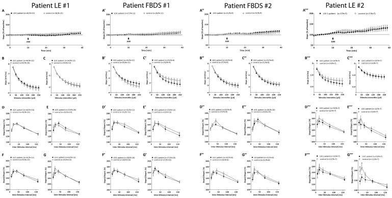
Figure 3.
Additional electrophysiological recordings. Field excitatory postsynaptic potentials (fEPSPs) of acute hippocampal slices of C57Bl/6-GFP mice treated with control (open circles) or with LGI1 IgGs of each patient (black circles, frequency of 0.1 Hz). (A–A’’’) fEPSP recording with 40% of maximum slope for 10 min. Continuous wash-in of LGI1 IgGs did not alter the fEPSP signal. Input–Output curves revealed no differences in postsynaptic function after 1 h (B–B’’’) and additionally after 6 h (C–C’’’) treatment of acute slices with either control or LGI1 IgG. PPF paradigm before (D–D’’’) and after (E’’’) the first LTP recording revealed no differences in presynaptic function after 1 h treatment of acute slices with either control or LGI1 IgGs. PPF paradigm before (F–F’’’) and after (G–G’’’) the second LTP recording after 6 h of ab treatment revealed no differences, despite a significantly lower PPF curve following 6 h of LGI1 IgGs treatment of patient LE#2 after the LTP measurement ((G’’’), p(10 ms) = 0.04, p(20 ms) = 0.04).

