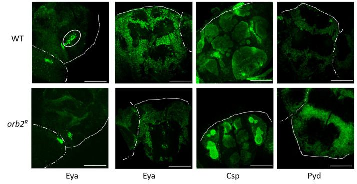
Figure 7.
Whole-mount staining of the Drosophila brain in WT and orb2R flies. Staining was performed with antibodies against the Eya, Csp, and Pyd proteins. A dashed line shows the board between the optic lobe and the central brain, and a solid line shows the board of the central brain. White ovals show a group of cells whose nuclei were stained with the antibody against the Eya protein in WT. For each antibody staining, the same exposure was used for samples of WT and Orb2R flies.

