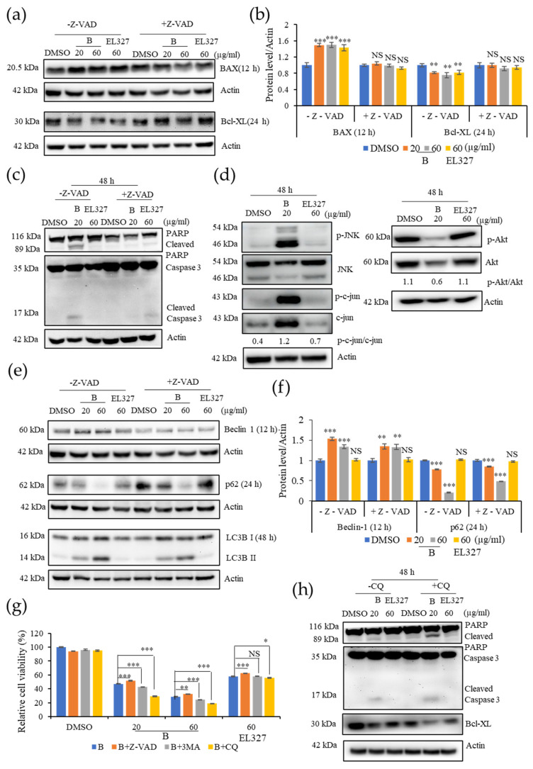
Figure 4.
Inhibition of B induced autophagy increases apoptosis in Caco2 cells. (a) Western blot analysis of the pro-apoptotic protein BAX and the anti-apoptotic protein Bcl-xL treated by B (20 or 60 µg/mL) or EL000327 (60 µg/mL) for 12 or 24 h in the presence or absence of Z-VAD-FMK (10 µM). (b) Quantification of BAX and Bcl-XL protein expressions. (c) Western blot of apoptotic proteins; PARP, Caspase-3treated by B (20 or 60 µg/mL) or EL000327 (60 µg/mL) for 48 h in the presence or absence of Z-VAD-FMK. (d) Expressions of the apoptotic signaling pathway related proteins p-JNK, JNK, p-c-jun, c-jun, p-AKT, and AKT in Caco2 cells treated with B (20 µg/mL) or EL000327 (60 µg/mL) for 48 h, as analyzed by Western blotting. (e) Western blot of autophagy related proteins; Beclin 1 (12 h), p62 (24 h), and LC3BI/II (48 h) in Caco2 cells pre-incubated with or without Z-VAD-FMK, and treated with B (20, 60 µg/mL) or EL000327 (60 µg/mL). (f) Quantification of Beclin 1 and p62 protein expressions. (g) The relative percentage cell viability of Caco2 cells treated with B (20, 60 µg/mL) or EL000327 (60 µg/mL) for 48 h, with or without Z-VAD-FMK (10 µM) and autophagy inhibitors 3 MA (1 mM) and CQ (10 µM). (h) Expression levels of PARP, caspase-3, and Bcl-xL determined by Western blot analysis after treatment with B (20, µg/mL) or EL000327 (60 µg/mL) for 48 h in the presence or absence of CQ (10 µM). Data represent the mean ± S.D. * p < 0.05, ** p < 0.01, *** p < 0.001; NS: no significant difference (p > 0.05), compared with the Z-VAD-FMK, 3MA, and CQ treated groups or the DMSO-treated control.

