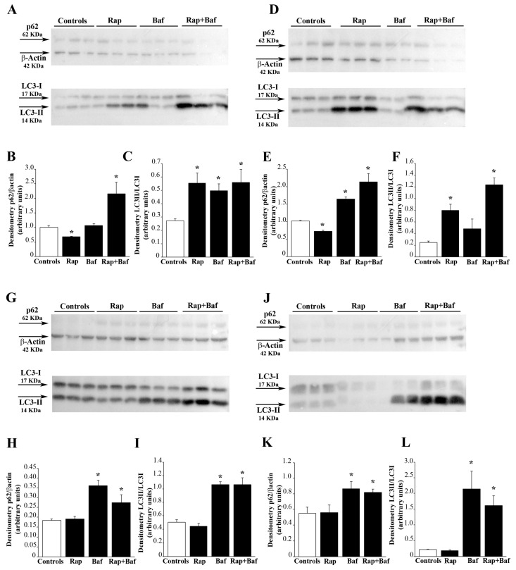
Figure 2.
Rapamycin treatment increases the autophagy flux. Western blot analysis of p62 and LC3-II in U87MG cells treated with 10 nM rapamycin for (A) 24 h and (D) 4 d, (G) 7 d and (J) 14 d after its removal. In some samples bafilomycin A1 (100 nM) was added during the last 3 h in both untreated and rapamycin-treated cultures. Densitometry analysis of the level of p62 compared with the housekeeping β-actin and LC3-II compared with LC3-I is also reported in the graphs. In detail, densitometry of p62/β-actin in cells treated with rapamycin for (B) 24 h and (E) 4 d, (H) 7 d and (K) 14 d after its removal is reported. Similarly, densitometry of LC3-II/LC3-I in cells treated with rapamycin for (C) 24 h and (F) 4 d, (I) 7 d and (L) 14 d after its removal is shown. Values are given as the mean ± SEM from three samples per experimental group. * p < 0.05 compared with controls.

