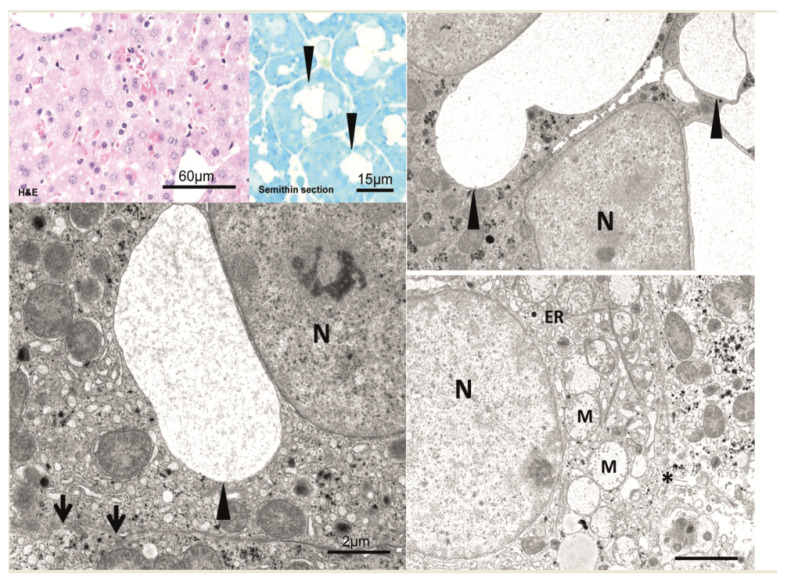
Figure 4.
After 10 to 20 min, vesicles increase in size and number, often forming vacuoles (arrowheads). Vacuoles can usually be found in proximity to the nucleus (N). They are optically empty and contain no electron-dense material. Vesicle-containing hepatocytes show intact cell borders (arrow). In contrast, some but other hepatocytes reveal signs of cell damage, i.e., swollen mitochondria (M), dilated endoplasmic reticulum (ER), and disrupted plasma membranes (asterisk). All findings were similar after the injection of the empty vector and the saline solution as well. Scale bars are indicated in the respective image.

