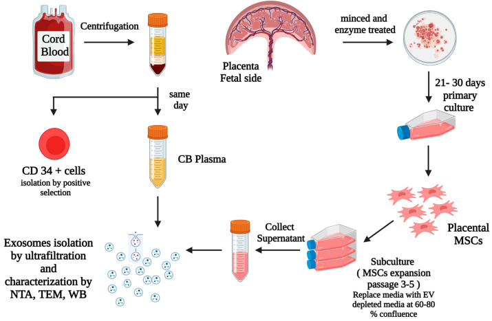
Figure 1.
This diagram summarizes our methods and the steps for the isolation of CD34+ cells via positive selection from UCB, isolation, processing, and culture of placental tissues from the fetal side, isolation of primary MSCs (passage 0), expansion of MSCs via subculture from primary MSCs to passage 3–5, replacement of the culture media with EV depleted media when the expanded cells were at 60–80% confluence, then collection of the MSCs culture supernatant after 48 h and exosomes isolation from CBP and MSCs culture supernatants. Created with BioRender.com.

