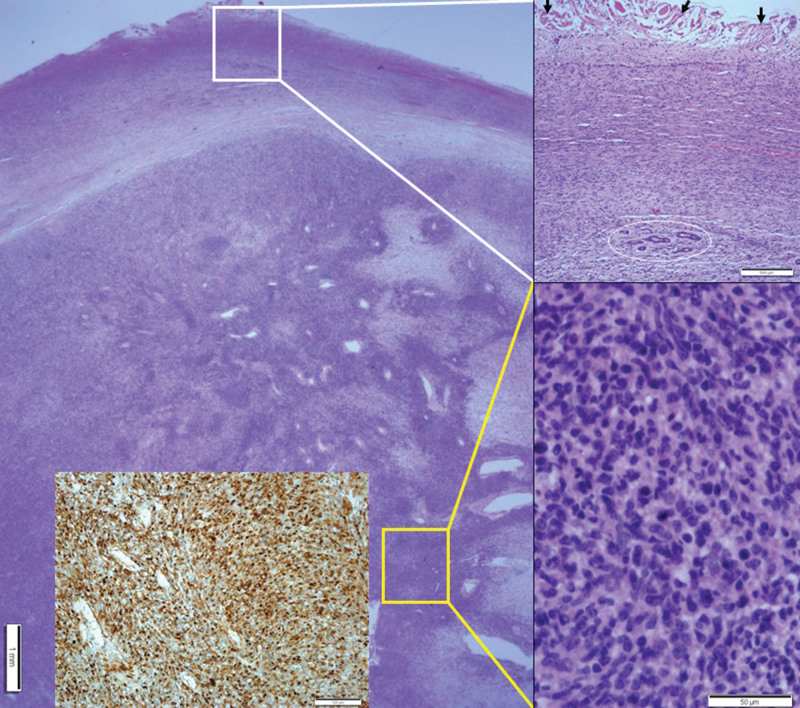
Figure 4.
Histopathologic findings of cutaneous MPNST. A well-circumscribed nodular mass showing dense cellular fascicles alternate with myxoid regions from the dermis to the deep subcutaneous layer. At a higher magnification, the section in the white box was found to have less cellular spindle cells with no cellular atypia. Entrapped skin appendages were identified in the circle. Arrows indicate dermal collagen at the resection margin. At a higher magnification, the section in the yellow box was found to have more cellular spindle cells with cellular atypia and occasional atypical mitoses. Inset: Positive immunohistochemical staining for the S-100 protein. MPNST = malignant peripheral nerve sheath tumor.

