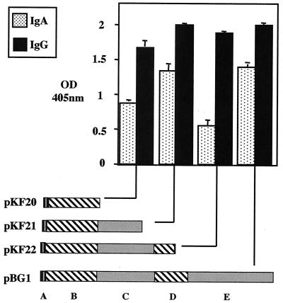
FIG. 6.
Graph representing ELISA results for the immunoglobulin binding by the respective indicated subclones, pKF20 to -22, expressing truncated SibA. The domains expressed by the subclones are a predicted coiled-coil region containing the bZip-like domain (A), a region of low complexity (B), predicted coiled-coil region 2 (C), and a proline-rich extended-sheet domain (D). The optical density (OD) (405 nm) is indicated on the vertical axis. Graphs were blanked against a PBS control which had no binding protein added. Error bars indicate standard deviations.

