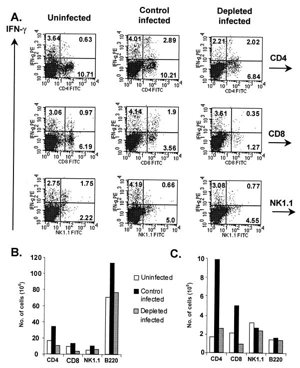
FIG. 5.
Neutrophil-depleted mice display decreased numbers of IFN-γ+ T cells during infection. (A) IFN-γ expression in CD4+, CD8+, B220+ and NK1.1+ subsets at 6 days postinfection. Numbers in quadrants reflect the percentages of cells. PE, phycoerythrin; FITC, fluorescein isothiocyanate. (B) Total numbers of cells in each subset. (C) Numbers of IFN-γ+ cells in each subset. Splenocytes were obtained and stimulated ex vivo before staining as described in Materials and Methods. Cells were then analyzed by flow cytometry. To obtain absolute cell numbers, percentages in each population were multiplied by the mean total cell number in corresponding spleens (three mice per group). Results represent three separate experiments.

