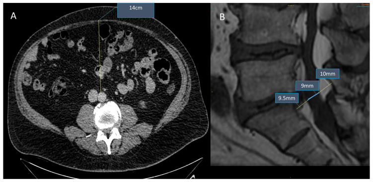
Figure 1.
Obese patient (45 years old, male, BMI = 31 kg/m2, dyslipidemia). Association between SEL and visceral fat deposition: CT of the abdomen (Panel A) reveals increased visceral fat deposition (AP diameter 14 cm), while lumbosacral MRI sagittal T1w (Panel B) reveals SEL grade 2: moderate (dural-sac/epidural fat index = 0.46).

