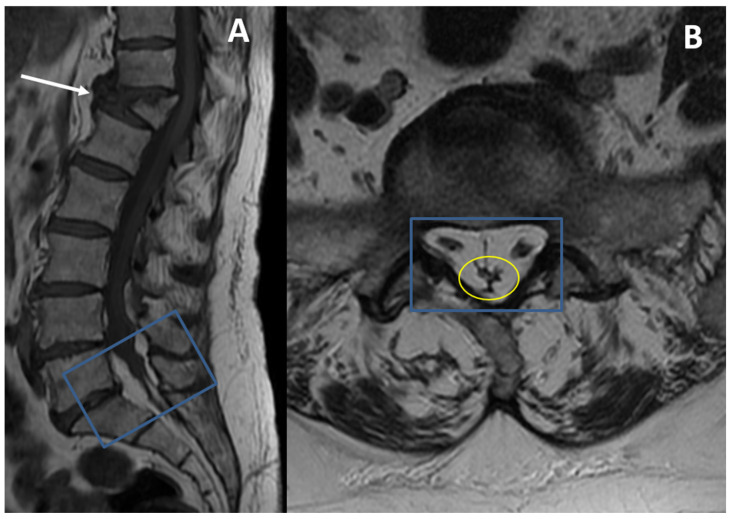
Figure 2.
A 72-year-old woman affected by dyslipidemia, obesity, and high blood pressure underwent follow-up MRI control for a vertebral fracture (T12); patient complained about chronic lumbar pain, which worsened after the fracture, and bilateral sciatica. (A) Sagittal T1-weighted imaging (WI); (B) axial T1-WI. A marked vertebral body collapse of the T12 vertebra is detectable (white arrow). Moreover, a condition of SEL at L5-S1 level (blue rectangles) with compression of the thecal sac resembling the letter “Y” (yellow oval) is found.

