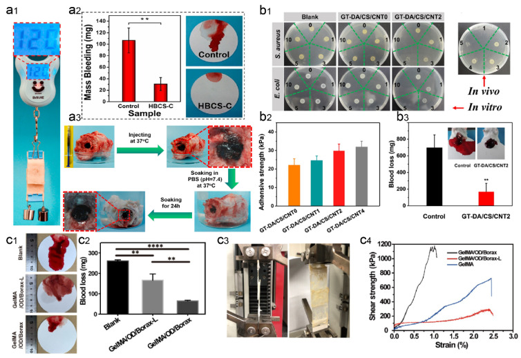
Figure 8.
Wet-adhesive hemostatic hydrogel in liver wounds. (a) Tissue adhesion and hemostatic effect of HBCS-C hydrogel. Reprinted (adapted) with permission from [100]. Copyright {2020} American Chemical Society. (a1) The images show good adhesion of HBCS-C hydrogel on porcine skin (adhesion area of 20 mm × 20 mm and heavy load of 120 g). (a2) Total blood loss from liver wounds in hydrogel-treated and untreated rats. (a3) Photographs showing the wet bioadhesive behavior and stability of HBCS-C hydrogels in an aqueous environment at 37 °C. (b) Antimicrobial, adhesion, and hemostatic properties of GT-DA/CS/CNT hydrogel. Figure modified from [102] with permission. (b1) In vitro antibacterial activity of hydrogels induced by NIR illumination, 0, 1, 3, 5, and 10 represent different irradiation times (min). (b2) Adhesion strength of the hydrogels after 1 h in the air before testing. (b3) Hemostatic ability of GT-DA/CS/CNT hydrogels. (c) Adhesion and hemostatic properties of GelMA/oxidized dextran/Borax hydrogels. Figure modified from [101] with permission. (c1) Photographs of liver blood loss after different treatments. (c2) Blood loss during hemostasis of the liver. (c3) Pictures of the modified test method used for the overlapping shear test. (c4) Strain–stress curves for the overlapping shear test. (** p < 0.01, **** p < 0.001).

