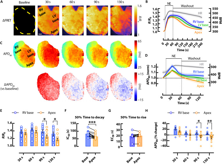
Fig. 3. Effects of β-adrenergic stimulation on cAMP responsiveness and APD heterogeneity in the mouse heart.
(A) Representative CFP/YFP whole-heart ∆FRET ratio images showing the spatiotemporal kinetics of cAMP activity (scale bar, 1 mm) and (B) average (mean = solid lines; SEM = shadow) CFP/YFP FRET ratio traces in response to bolus of 1.5 μM NE from different regions of the heart (blue = RV base; green = LV base; orange = apex), with corresponding changes in HR (gray). (C) APD80 maps from the heart in (A) (top) with change in APD80 versus baseline (∆APD80) (bottom) and (D) average (mean = solid lines; SEM = shadow) normalized APD80 over time showing APD prolongation and then shortening after application of 1.5 μM NE, with corresponding changes in HR (gray). Mean scatter dot plots from RV base (blue) and apex (orange) regions following 1.5 μM bolus NE and washout showing; (E) FRET ratio responses at different time points; (F) FRET ratio 50% decay time calculated from half maximal inhibitory concentration (IC50); (G) FRET ratio 50% rise time calculated from median effective concentration (EC50); and (H) APD80 % changes from baseline (time 0). N = 13 hearts from 6 male and 7 female mice. Representative heart in (A) and (C) from a female mouse. *P < 0.05; **P < 0.01, by two-way ANOVA with multiple pairwise comparisons; ***P < 0.001, by two-tailed paired t test.

