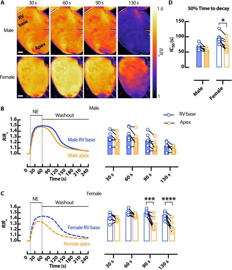
Fig. 4. Sex-dependent differences in cAMP spatiotemporal responses in the mouse heart.
(A) Representative ∆FRET ratio images showing the spatiotemporal kinetics of cAMP activity in male (top) and female (bottom) CAMPER hearts after application of 1.5 μM NE. Scale bars, 1 mm. Corresponding representative FRET ratio traces and mean scatter dot plots from RV basal (blue) and apical (orange) regions following 1.5 μM bolus NE at different time points in male (solid) (B) and female (dashed) (C) CAMPER mouse hearts. (D) 50% time to decay calculated from FRET ratio IC50. N = 6 male and 7 female hearts. *P < 0.05; ***P < 0.001; ****P < 0.0001, by two-way ANOVA with multiple pairwise comparisons.

