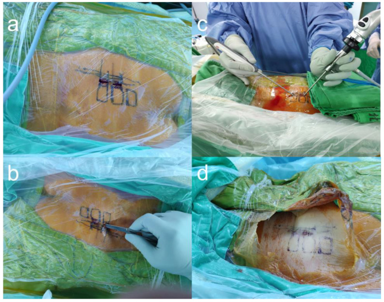
Figure 1.
The entry points and working channels of biportal endoscopic radiofrequency ablation. (a) Under fluoroscope, the S1–S3 foramina were marked on the skin. (b) Two incisions were made, one near the S1 foramen and another near the S2 foramen. (c) One foramen was for the endoscopic channel, and the other was for the insertion of the ablation wand. (d) Sutured wound after the surgery.

