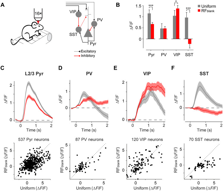
Fig. 4. Cell-specific responses to contextual stimuli in V1.
(A) We recorded cell-specific Ca2+ responses of V1 neurons using two-photon microscopy (left). Schematic of V1 microcircuit involving pyramidal and the three main classes of interneurons (right). (B) Average activity evoked by full-screen grating (uniform, gray) or contextual (RFblank, red) stimuli in a time window of 0 to 2 s in 537 pyramidal neurons (Pyr), 87 PV neurons, 120 VIP neurons, and 70 SST neurons. ***P < 0.001 and *P < 0.05, LME comparing response to uniform and RFblank stimulus. (C) Average time course of activity across 537 L2/3 pyramidal neurons in V1 in five mice evoked by the uniform (gray) and RFblank (red) stimuli (top). Activity of individual L2/3 neurons in a time window of 0 to 2 s (bottom). (D) Average activity of 87 PV neurons in V1 of four PV-Cre mice. (E) Average activity of 120 VIP neurons in V1 of four VIP-Cre mice. (F) Average activity of 70 SST neurons in V1 of five SST-Cre mice.

