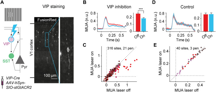
Fig. 5. Involvement of disinhibitory circuit in the contextual responses in V1.
(A) We optogenetically inhibited VIP neurons in eight VIP-Cre mice using cell-specific expression of the inhibitory opsin stGtACR2, while electrophysiologically recording V1 activity. Left: Schematic of the disinhibitory circuit involving VIP and SST neurons. Right: Image of coronal section of V1 spanning from the bottom to the top of the cortex. Expression of the optogenetic construct is visualized by the reporter molecule FusionRed. (B) Average, normalized multi-unit activity (MUA) across 316 V1 recording sites evoked by the RFblank stimulus, with (blue) and without (red) inhibition of VIP neurons (left). Average MUA was reduced when VIP neurons were inhibited (***P < 0.001, LME; time window, 0 to 0.5 s) (right). (C) Activity of V1 recording sites from 0 to 0.5 s after presentation of the RFblank stimulus with (y axis) and without (x axis) inhibition of VIP neurons. Different colors represent different electrode penetrations. (D and E) Data from control experiment without the optogenetic construct in VIP neurons and identical laser intensities. Laser light did not affect the response to the RFblank stimulus.

