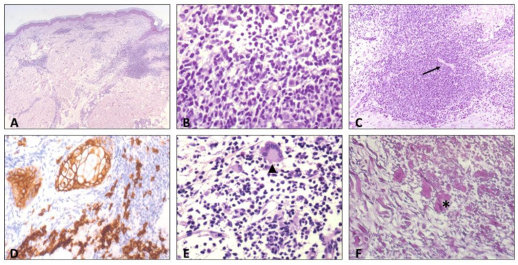
Figure 3.
Histologic findings: (A) superficial and deep dermal perivascular inflammatory infiltrate, hematoxylin eosin (HE). (B) High-power view of the inflammatory infiltrate, showing predominance of lymphocytes and plasma cells, HE. (C) Endothelial swelling of dermal blood vessels (arrow), HE. (D) Immunostaining for CD138 highlights plasma cells infiltrate, (E) scattered multinucleated giant cells in the infiltrate, also of the Langhans type (arrowhead), and (F) elastophagocytosis (asterisk), Weigert-Von Gieson staining.

