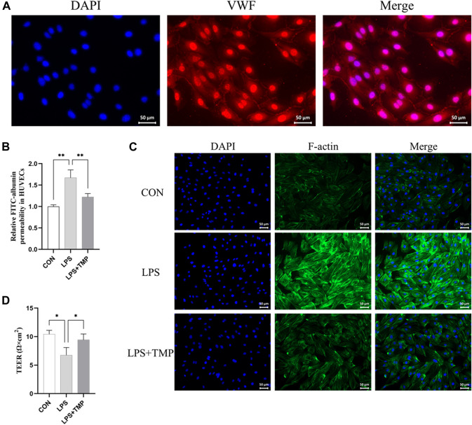
FIGURE 5.
Effects of TMP on the cytoskeleton in vitro. (A) VWF staining was used to identify endothelial cells (scale bar = 50 μm). (B) Transwell chamber FITC albumin was used to determine the effect of TMP (15 ng/ml) on HUVEC after 12 h of LPS (200 ng/ml) exposure. (C) Immunofluorescence assay for the cytoskeleton (scale bar = 50 μm). (D) Transendothelial electrical resistance (TEER) in TMP-treated, untreated, and control HUVECs. * p < 0.05, ** p < 0.01, n = 3 per group.

