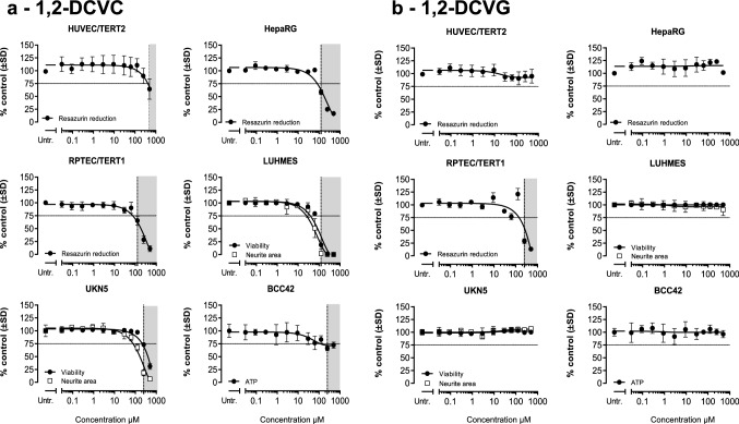
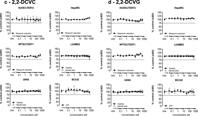
Fig. 3.
Cell viability assessment after DCVGs and DCVCs exposure. Measurements were performed after 24 h exposure of eleven concentrations (0.03, 0.09, 0.29, 0.9, 2.9, 9.3, 29.7, 62.5, 125, 250, 500) of indicated TCE conjugates in the six human cell models tested. Viability in HUVEC/TERT2, HepaRG and RPTEC/TERT1 was assessed by resazurin reduction. Viability and neurite area in LUHMES and UKN5 were assessed with Calcein-AM and H-33342 staining. ATP content was used as viability parameter in BCC42 cells. Values represent the mean % medium control ± SD for each different assay. Untr: untreated samples were used as negative controls. a 1,2-DCVC, b 1,2-DCVG, c 2,2-DCVC, d 2,2-DCVG


