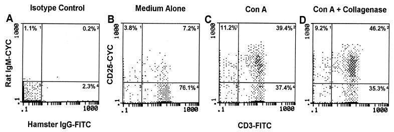
FIG. 1.
Expression of CD25 following ConA stimulation and collagenase type IV treatment. LNC from four mice were cultured alone (B) or in the presence of ConA (C) for 24 h. Following the culture period, ConA-stimulated cells were incubated with or without collagenase (D) for 45 min. At the conclusion of these treatments, the cells were dually labeled with anti-CD3 and anti-CD25 antibodies or isotype control antibodies (A) and were analyzed by flow cytometry using isotype control antibodies to set fluorescent limits (gates). Numbers within each quadrant represent the percentage of fluorescent positive cells within lymphoid cell limits. Data shown are representative of three separate experiments. FITC, fluorescein isothiocyanate; CYC, cytochrome c.

