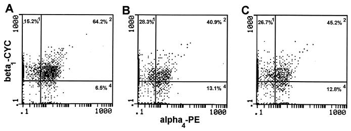
FIG. 4.
Expression of α4β1 on CD3+ LNC during experimental vaginal candidiasis. Lymphocytes were isolated from the draining lumbar lymph nodes of uninfected mice (A), mice with primary infection (B), and mice with secondary infection (C) on day 10 postinoculation. The LNC were triply labeled with anti-CD3, anti-α4, and anti-β1 antibodies and analyzed by flow cytometry. The numbers within each histogram represent the percentage of gated CD3+ cells present within each quadrant. PE, phycoerythrin; CYC, cytochrome c.

